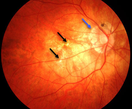
OCT scan of the top (A) and bottom (B) macula showing ERM, myopic foveoschisis with associated lamellar macular hole OS>OD and thin choroid. Posterior vitreous detachment and 2+ floaters were observed in both eyes.įig 2. Optic nerve heads (ONH) had a myopic tilt, extensive peripapillary atrophy temporally and a cup-to-disc ratio of 0.15 right eye and 0.25 left eye without evidence of clinic nerve fiber layer defects. Additionally, the tessellate, tigroid appearing fundus had myopic pigment changes with no foveal light reflex. There was a small amount of subretinal fluid underlying the retinal tears that did not extend beyond the newer laser scars, otherwise there was no new breaks or detachments with scleral indentation performed 360 in each eye. The previously reported posterior pole findings appreciated via dilated fundoscopy were as follows: treated retinal tear with old cryotherapy and new barrier laser scars (12:30) in the right eye, and treated retinal tears (11:00, 1:30) with pigmented lattice degeneration (5:00-6:00), all surrounded by barrier laser in the left eye. Evaluation of the anterior segment revealed bilateral trace nuclear sclerosis and 2+ posterior subcapsular cataracts. Intraocular pressure (IOP) was 20mmHg OD and 21mmHg OS via Goldmann applanation tonometry. Extraocular motilities, confrontation visual fields and pupils were all unremarkable. Click image to enlarge.īest corrected distance visual acuity (BCVA) was 20/30-2 OD, 20/40+1 OS, 20/25-2 OU, PHNI at distance and 20/25 OU at near with progressive lens prescription of -14.50 2.00x008 OD and -13.00-2.00x170 OS, +2.25 add OU. Laser scars surrounding a laser tears superior OD/OS and lattice degeneration inferior OS. UWF retinal photo of the top (A) and bottom (B) eye showing a tessellated fundus, tilted nerve with temporal crescent. She had no remarkable medical history she was not taking any medications and had no known drug allergies. She had documented bilateral myopic maculopathy with epiretinal membranes and right eye foveoschisis. She underwent barrier laser retinopexy of retinal tears in both eyes approximately twelve years prior with re-treatment ten years later for additional laser barricade around the retinal tears in addition to inferior lattice degeneration in her left eye. The patient had an extensive ocular history with regards to retinal changes related to PM. She had yellow filter glasses that provided relief. Case PresentationĪ 58-year-old Native American female presented for a routine eye examination with complaints of floaters and glare out of both eyes that she had experienced consistently for many years. This case report will discuss a female patient with pathologic myopia and outlines the various manifestations and management options. This case report outlines the various manifestations and management options of PM.

Moreover, identifying myopia at an early age and employing interventions to suppress progression to PM should become a top priority. The prognosis of PM varies depending on the clinical features thus, it is critical to identify and monitor high risk patients, appreciate the severity, and take a collaborative approach pursuing interventions for vision threatening sequelae. Excessive axial length elongation drives biomechanical stretching of the posterior globe often associated with retinal and choroidal changes that inevitably lead to severe and irreversible visual impairment. Pathologic myopia (PM) represents a spectrum of degenerative ocular structural complications that arise secondary to high myopia. The contest is sponsored by Topcon, Visionix (Optovue) and Heidelberg. This case, selected by the ORS Awards Committee, was co-winner of the sixth annual Larry Alexander Resident Case Report Contest. That group chose to honor his legacy by accepting case reports from optometric residents across the country relating to vitreoretinal disease. Alexander was a past president of the Optometric Retina Society (ORS). In addition to being an optometric physician, author and educator at the University of Alabama Birmingham School of Optometry, Dr.

In April 2016, optometry lost a giant when the author of the seminal work Primary Care of the Posterior Segment, Larry Alexander, OD, died.


 0 kommentar(er)
